
Cancer’s most dangerous habit is not growth but travel. A tumor that stays put can often be removed; a tumor that learns to send pieces of itself into the bloodstream and lymph system is a different kind of problem. For decades, researchers have studied how cancer cells communicate with their surroundings, and one of the most active areas of inquiry concerns extracellular vesicles—tiny, bubble-like structures that cells release and that can carry proteins, RNA, and DNA to distant locations. Now, a team at McGill University Health Centre has discovered a previously unknown type of vesicle, one that appears specifically tied to cancer cell migration and carries something remarkable: the entire genome of the parent cell.
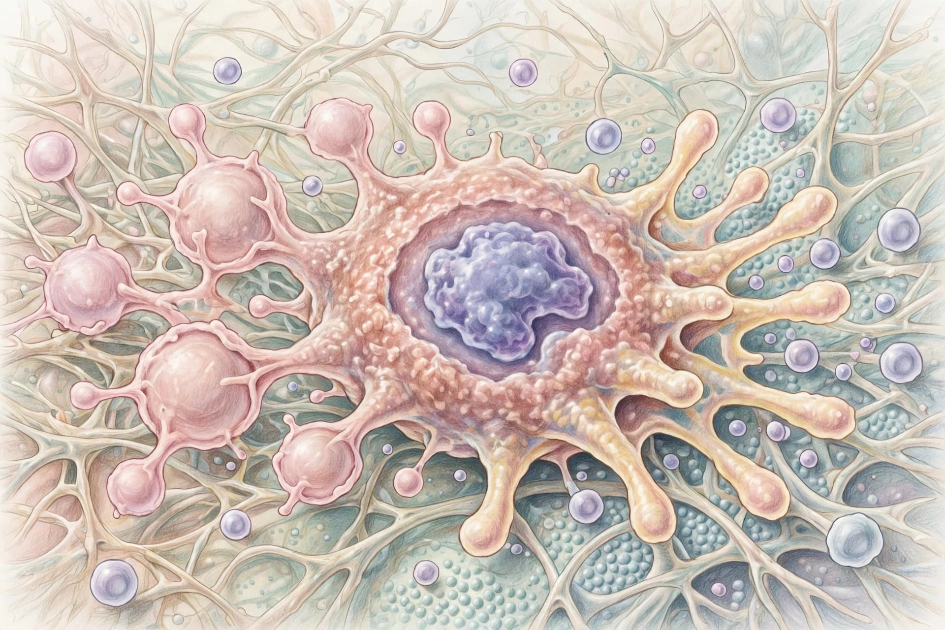
The vesicles are called MoVes, short for motile vesicles. They are large by vesicle standards, ranging from two to five micrometers, and they are formed at the trailing edge of migrating cancer cells through a process distinct from other known vesicle pathways . Live-cell imaging and scanning electron microscopy revealed that MoVes are actively shed by cancer cells but not by non-transformed cells, and their production is linked to the epithelial-to-mesenchymal transition, or EMT—a process that allows cells that normally stick together to become mobile and invasive .
What makes MoVes particularly striking is their cargo. Whole-genome sequencing demonstrated that the DNA inside MoVes spans the genome of the parent cell, recapitulating its mutational landscape . This is not a fragment or a subset; it is, in effect, a copy of the cancer cell’s genetic identity, packaged into a membrane-bound structure and released into the extracellular space. The vesicles also carry functional organelles, including mitochondria, and proteomic profiling identified enrichment of FAM3C, a protein associated with EMT, along with other proteins involved in organelle biology and oxidative stress response .
The discovery was, in a sense, accidental. The McGill team was not looking for a new vesicle type. They were trying to understand how cancer cells release DNA, and in the process they uncovered a biological mechanism no one had described before. “What makes this discovery particularly exciting is that we were not looking for a new type of vesicle,” said Thupten Tsering, the first author of the study . The work, published in Nature Cell Biology, was led by Prof. Julia Burnier, a scientist in the Cancer Research Program at the Research Institute of the MUHC.
Functional assays added another layer to the story. MoVes-associated mitochondrial cargo was found to enhance oxidative phosphorylation in recipient cells—meaning the vesicles can deliver not just genetic information but metabolic capability . The researchers also observed that aggressive triple-negative breast cancer cells produced far more MoVes than non-cancerous breast cells, and that migration-inhibitory drugs significantly reduced their biogenesis . In animal models, MoVes were most abundant in the liver, suggesting a possible role in the early stages of metastatic spread .
The implications are still being worked out. If MoVes carry the full genomic landscape of a tumor, they could potentially serve as biomarkers—measurable indicators that might help detect cancer, monitor its progression, or assess treatment response. The Burnier lab is now working to determine whether MoVes can be detected in patients, and to understand the mechanisms that control their formation and release . There is also the question of what recipient cells do with the DNA and organelles they receive. Do they incorporate the genetic material? Does it alter their behavior in ways that favor metastasis?
These are early questions, and the answers are not yet clear. But the discovery adds a new thread to the tangled story of how cancer spreads. Extracellular vesicles have long been suspected of playing a role in preparing distant tissues for metastasis, creating what some researchers call a “pre-metastatic niche.” MoVes, with their full genomic cargo and their specific association with migration, may be a particularly direct form of that communication—a cancer cell sending not just a signal but a complete copy of itself into the world.
For patients, the hope is distant but real. A better understanding of how cancer cells shed their DNA and organelles could reveal new targets for therapy, new ways to detect tumors earlier, or new strategies to interrupt the processes that allow cancer to travel. The road from a discovery like this to a clinical tool is long, and most such discoveries do not make it. But the first step is always the same: seeing something that was not seen before.
Image note: The images accompanying this article are AI-generated illustrations and do not depict actual cells or laboratory results.
Sources: McGill University Health Centre, Nature Cell Biology, BioStudies (EMBL-EBI), ProteomeXchange
Published by Banx Network. This article is part of the Banx decentralized media programme, powered by the BXE token on the XRP Ledger.