
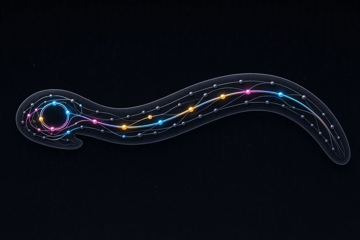
In the microscopic world of the nematode Caenorhabditis elegans, simplicity holds the key to complexity. With only 302 neurons, this tiny worm possesses a nervous system that is fully mapped, offering scientists a unique window into how brain structure gives rise to behavior. Recent research has focused on the synchronization of neural activity within this compact network, exploring how specific circuits coordinate to produce smooth, rhythmic movements. The findings shed light on the fundamental principles of neuronal connectivity and functional integration.
The study examines how groups of neurons fire in unison to control locomotion, a behavior that requires precise timing and coordination. By combining detailed anatomical maps with functional imaging, researchers have identified specific patterns of connectivity that facilitate this synchronization. These patterns are not random; they reflect an optimized architecture that allows the worm to navigate its environment efficiently, responding to stimuli with agility and grace.
Synchronization is a critical feature of neural systems, enabling disparate parts of the brain to work together as a cohesive whole. In C. elegans, this process is driven by both electrical and chemical synapses, which transmit signals between neurons. The balance between these types of connections determines the stability and flexibility of the network, allowing the worm to switch between different behavioral states, such as forward movement, reversal, or turning.
Understanding these mechanisms in a simple organism provides insights that may apply to more complex brains, including our own. Many neurological disorders, such as epilepsy and Parkinson’s disease, involve disruptions in neural synchronization. By studying how C. elegans maintains healthy coordination, scientists can identify potential targets for therapeutic interventions in humans, bridging the gap between basic biology and clinical medicine.
The research also highlights the importance of structural connectivity in shaping function. The physical layout of the neural network constrains the possible patterns of activity, guiding the flow of information. This structure-function relationship is a central theme in neuroscience, and C. elegans serves as an ideal model for testing theories about how anatomy dictates behavior. The transparency of the worm allows for real-time observation of neural activity, providing unprecedented detail.
Advanced computational models play a crucial role in this analysis, simulating the dynamics of the neural network based on known connectivity data. These models help predict how changes in synaptic strength or neuron properties affect overall behavior. By comparing simulation results with experimental observations, researchers can refine their understanding of the underlying principles, creating a feedback loop of discovery and validation.
The elegance of the C. elegans system lies in its completeness. Every neuron and connection is known, allowing for a holistic approach to studying brain function. This comprehensive view is rare in neuroscience, where most organisms are too complex to map entirely. As a result, findings from C. elegans often serve as foundational truths that inform broader biological concepts.
As we continue to unravel the secrets of this tiny worm, we gain a deeper appreciation for the sophistication of even the simplest life forms. The synchronization of its neurons is a testament to the efficiency of evolution, demonstrating how complex behaviors can emerge from minimal components. In the rhythm of its movement, we see the universal language of life.
AI Image Disclaimer: The visual aids included here are AI-generated illustrations of neuronal networks and nematode anatomy, not actual microscopy images or functional scans.
Sources: eLife Journal of Neuroscience WormBook
Published by Banx Network. This article is part of the Banx decentralized media programme, powered by the BXE token on the XRP Ledger.