

Key Highlights
- New Technique:Researchers at University College London developed a method to visualize bacterial poles.
- Study Findings:The poles reveal critical information about bacterial activity and antibiotic resistance.
- Publication:The research was published in the journal Nature Communications.
[New Imaging Technique Unveils Bacterial Secrets]
For decades, scientists have struggled to fully understand the structure of bacteria, particularly the poles where cells divide. These regions, crucial for understanding how bacteria grow and how some antibiotics work, have remained elusive. However, a groundbreaking study from University College London has introduced a new technique that allows researchers to visualize these poles in unprecedented detail.
The innovative method involves coating a glass surface with a chemical that binds to proteins on the bacterial surface, effectively anchoring the cells in place while allowing them to grow and reproduce. Initially, the bacteria lay flat, but after about fifty minutes, many stood upright, exposing their poles to an atomic force microscope. This advanced imaging technique taps gently across the cell surface, creating high-resolution images that surpass traditional light microscopy.
What the researchers discovered at the poles was surprising. Instead of smooth, uniform structures, they found evidence of cellular activity. Bacteria that were rapidly dividing displayed fold-like defects at their poles, indicating that they had not fully remodeled after division. In contrast, less active bacteria exhibited more rounded poles. This suggests that the poles are not merely passive structures; they are dynamic sites of ongoing construction and reconstruction, reflecting the cell's history and current state.
The study, published in the prestigious journal Nature Communications, also highlighted significant differences between bacterial species. For instance, E. coli was found to have a network of tiny pores on its surface, while pathogens like Pseudomonas aeruginosa and Acinetobacter baumannii lacked this feature entirely. These distinctions are crucial, as they imply that the outer structures of these bacteria are constructed differently, which could affect how antibiotics interact with them. Understanding these differences is vital for addressing the growing issue of antibiotic resistance.
Professor Bart Hoogenboom, the senior author of the study, likened the achievement to "opening a view of the back of the moon." He emphasized the novelty and importance of imaging bacterial poles, which may serve as specific targets for antibiotic treatments. Some antibiotics appear to preferentially attack these regions, making this research particularly relevant for developing new strategies against drug-resistant infections.
While this work is fundamental science and does not immediately lead to new drugs or treatments, it lays the groundwork for future research. By elucidating the structure of bacterial surfaces at such a high resolution, scientists hope to identify new avenues for targeting drug-resistant strains and developing innovative methods to breach the protective barriers that shield these cells from antibiotics.
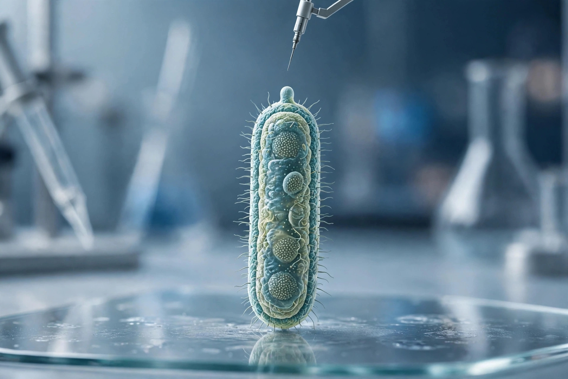
The images accompanying this article are AI-generated illustrations and do not represent actual microscopy images of bacterial cells.
Published by Banx Network. This article is part of the Banx decentralized media programme, powered by the BXE token on the XRP Ledger.