
There is a particular difficulty in the quiet work of diagnosis. Ovarian cancer, in its early stages, often announces nothing. No pain, no lump, no clear signal that something has gone wrong. It is a disease that prefers to remain unseen, and by the time it makes itself known, the window for easy intervention has often narrowed. This is why researchers continue to look for ways to see more clearly—to combine what imaging reveals with what blood can tell us, and to build tools that help clinicians make better decisions before surgery. A new study from China offers one such tool, and its early results are quietly encouraging .
The study, published in Scientific Reports, focused on epithelial ovarian tumors, the most common type of ovarian cancer. The researchers set out to develop and validate a predictive model that integrates magnetic resonance imaging (MRI) features with serum tumor markers—specifically CA125 and HE4—to distinguish benign from malignant tumors . The premise is straightforward but powerful: neither imaging nor blood tests alone are perfect, but together, they may offer a clearer picture. MRI can reveal the shape, structure, and diffusion characteristics of a tumor. Serum markers can hint at its biological behavior. A model that weighs both may see what either alone might miss.
The research team, based at Huzhou Maternity & Child Health Care Hospital, analyzed data from 222 patients as a training cohort and 102 patients from a separate hospital as an external validation cohort . This external validation is important. Many predictive models perform well in the data from which they are built but falter when applied to new patients in new settings. By testing their model on patients from a different institution, the researchers were able to assess whether it might hold up in real-world use. The results were notable. In the training cohort, the combined model achieved an area under the curve (AUC) of 0.965, with 94.4 percent sensitivity and 84.5 percent specificity. In the external validation cohort, the AUC was 0.972, with 90.9 percent sensitivity and 95.0 percent specificity .
The independent predictors identified by the model are not unfamiliar to clinicians. Tumor morphology—whether the mass appears cystic, solid, or mixed—was one. Diffusion-weighted imaging signal intensity, which reflects how freely water molecules move within tissue, was another. The presence of ascites, the accumulation of fluid in the abdomen, was a third. And the serum markers CA125 and HE4 completed the set . None of these is new to the diagnostic toolkit. What the study offers is a way of combining them, of assigning them weights derived from data rather than intuition, and of presenting a single risk estimate that can inform the next step in care.
The clinical implications are practical. Ovarian tumors are often evaluated surgically, and distinguishing benign from malignant before an operation can help avoid unnecessary procedures or, conversely, ensure that malignant tumors are treated with appropriate urgency. A model that can assist in this preoperative risk stratification—that can say, with reasonable confidence, that a mass is more likely to be benign or more likely to be malignant—could be a useful adjunct to clinical judgment . It is not a replacement for the physician's eye, nor for the pathologist's microscope. But it may help focus attention where it is most needed.
The study has limitations, as all studies do. It is retrospective, drawing on existing records rather than following patients prospectively. The sample size, while adequate for model development, is modest. The model was built and validated in Chinese populations, and its performance in other populations will need to be tested. These are not criticisms so much as invitations—calls for the kind of broader, multicenter, prospective research that can confirm and refine what has been found here . The researchers themselves note that future studies should focus on larger and more diverse cohorts.
Still, the work represents a step in a direction that many in the field are moving: toward integration, toward models that synthesize different kinds of information, and toward tools that are simple enough to be used in routine practice. A predictive model is not a cure. It does not prevent cancer, nor does it treat it. But it may help clinicians see more clearly, and in a disease that thrives on being unseen, that clarity has value.
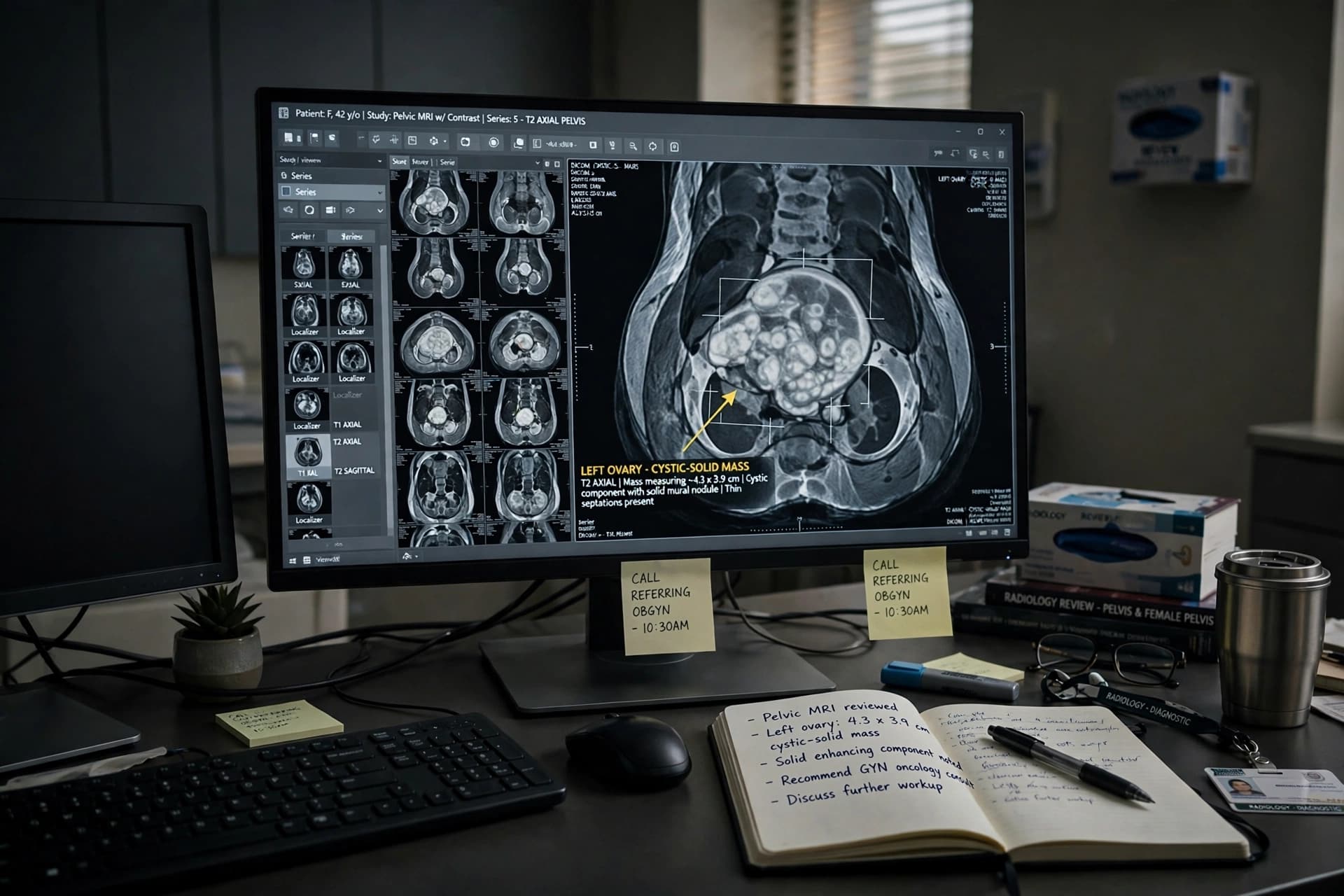
Image Disclaimer: The image accompanying this article was created with AI tools and is not a clinical photograph or medical scan.
Sources: Scientific Reports (Nature), Huzhou Maternity & Child Health Care Hospital, Sir Run Run Shaw Hospital
Published by Banx Network. This article is part of the Banx decentralized media programme, powered by the BXE token on the XRP Ledger.