
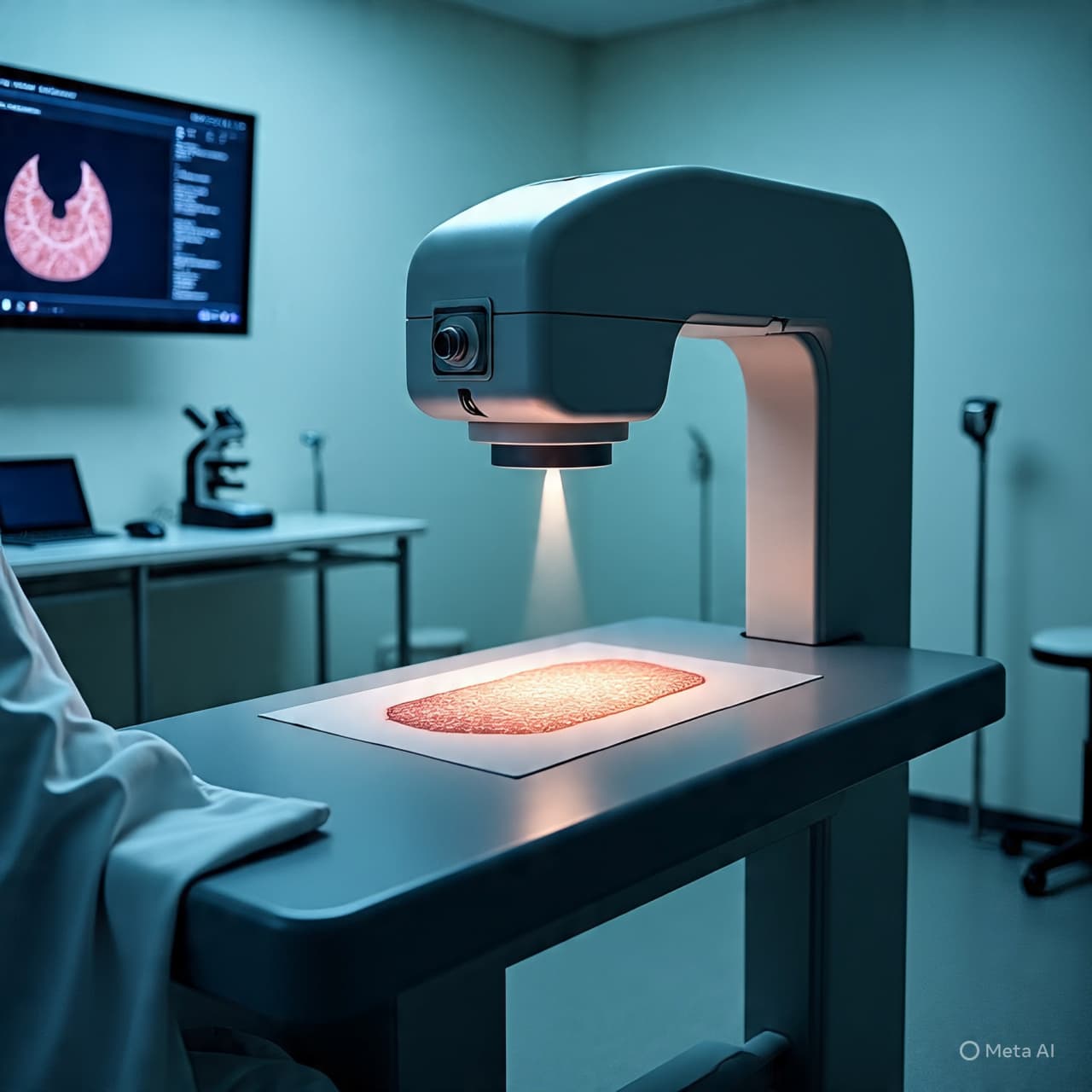
There are forms of seeing that do not rely on light as we know it, but on subtler frequencies that move through the world without drawing attention to themselves. In laboratories and clinical spaces, these invisible waves carry information quietly, revealing what lies beneath the surface without disturbance.
Among them are terahertz waves, occupying a region between microwave and infrared, long regarded as difficult to harness but rich with possibility. They pass through certain materials while reflecting off others, offering a way to distinguish between structures that might otherwise appear similar. For years, their potential in medical imaging has been recognized, though practical limitations have kept their use largely within controlled environments.
Now, a new imaging system has begun to shift that balance. By refining how terahertz signals are generated, captured, and processed, researchers have developed an approach that significantly reduces the time required to produce diagnostic images. What once demanded extended measurement and complex reconstruction can now be achieved more efficiently, bringing the technology closer to clinical application.
The implications unfold gradually. In medicine, time often shapes outcomes, particularly in contexts where early detection is critical. Faster imaging does not simply improve convenience; it alters the rhythm of diagnosis, allowing clinicians to move more quickly from observation to decision.
Terahertz imaging carries additional qualities that make it distinct. Unlike ionizing radiation, it operates at energy levels that do not damage tissue, offering a non-invasive alternative for examining biological structures. It is also sensitive to water content and molecular composition, enabling it to detect subtle differences between healthy and abnormal tissue.
In practice, this means that conditions such as skin cancers or tissue irregularities may be identified with greater clarity. The technology does not replace existing imaging methods, but adds another layer, one that can complement established tools by providing different kinds of information.
The new system achieves its speed through advances in both hardware and computation. Improved detectors capture signals more efficiently, while algorithms process the resulting data with greater speed and accuracy. The combination allows for near real-time imaging, a step that moves the technology beyond experimental use toward integration in clinical workflows.
There are, however, considerations that remain. The transition from laboratory to hospital involves not only technical refinement, but also validation, cost assessment, and adaptation to practical constraints. Each of these steps shapes how and when the technology might become widely available.
What emerges from this development is not a sudden transformation, but a steady progression. Terahertz imaging, once limited by its own complexity, begins to find a clearer path toward application. The process reflects a broader pattern in science, where advances often arrive not as singular breakthroughs, but as accumulations of refinement.
There is a certain quietness in this kind of progress. The waves themselves remain invisible, their movement undetected by the senses, yet their impact grows as they are brought into use. In the space between light and matter, a new form of vision takes shape, one that may alter how conditions are seen, and how early they can be understood.
Researchers have developed a faster terahertz imaging system that enables more efficient clinical diagnostics, improving the speed and potential accuracy of non-invasive medical imaging. The technology is moving closer to practical use in healthcare settings.
AI Image Disclaimer
Illustrations were created using AI tools and are not real photographs, but visual interpretations of the described technology.
Source Check Nature Science IEEE Spectrum Medical News Today Scientific American
Published by Banx Network. This article is part of the Banx decentralized media programme, powered by the BXE token on the XRP Ledger.