
Inside every living cell, there are moments that unfold without sound yet carry the weight of endings. A cell grows, divides, repairs itself, and moves through its quiet routines. But when the time comes — when damage becomes too great or signals demand change — the cell may follow a different path, one that leads deliberately toward its own conclusion.
Biologists call this process programmed cell death, or apoptosis. It is not an accident of biology but a carefully orchestrated act, one that allows organisms to remove damaged, infected, or unnecessary cells while preserving the health of the larger body.
For decades, scientists have studied the molecular signals that guide this process. Yet much of the story has unfolded at scales so small that even the most advanced imaging techniques reveal only fragments of the machinery involved.
Now researchers at the University of Michigan have uncovered a new structural detail within this hidden world — a formation of higher-order protein rings that appear to help regulate how programmed cell death begins.
The discovery focuses on proteins involved in the pathways that determine whether a cell will survive or initiate apoptosis. These proteins do not always act alone. Instead, they assemble into larger complexes, structures that allow signals to travel and amplify within the cell.
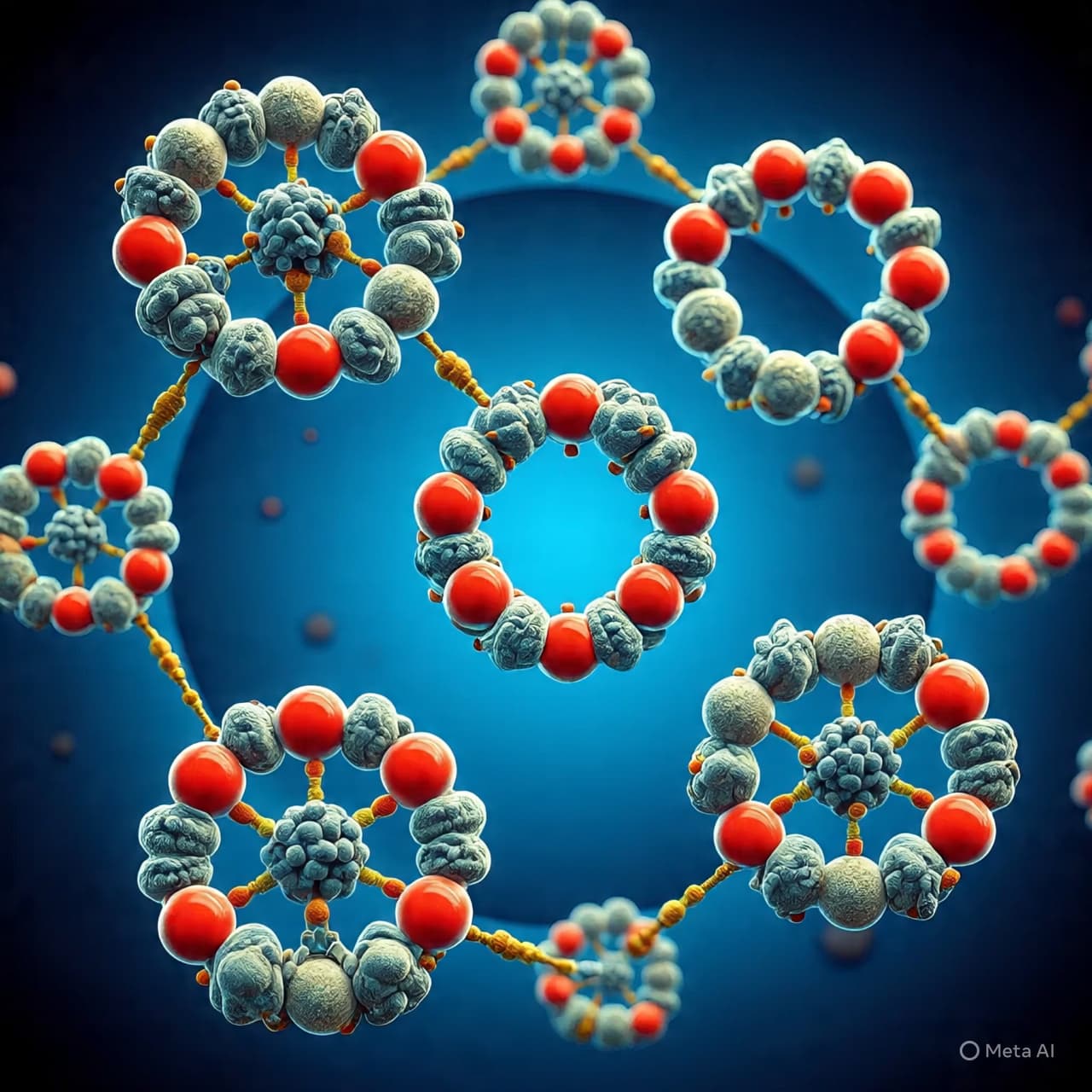
Using advanced structural biology techniques, the researchers observed that certain proteins form circular assemblies — rings composed of repeating molecular units that connect together in precise arrangements. These higher-order structures appear to organize the signaling machinery responsible for triggering cell death.
Rather than functioning as isolated molecules, the proteins assemble into coordinated frameworks that guide how the apoptotic signal spreads through the cell.
In the newly identified structures, the ring-shaped complexes create platforms where signaling proteins gather and interact. This architecture may help control when apoptosis begins and how strongly the signal propagates through the cellular environment.
Such organization matters because apoptosis must be tightly regulated. If cells die too readily, tissues can weaken and diseases such as neurodegeneration may arise. If cells fail to die when they should, damaged cells can accumulate, contributing to cancer and other disorders.
The study therefore adds an important piece to the puzzle of how cells maintain this delicate balance.
Researchers also note that higher-order protein assemblies — including rings, filaments, and lattices — are increasingly recognized as central elements in many biological processes. In immune responses, inflammation, and cell signaling, proteins often form large structures that act like scaffolding for complex biochemical reactions.
The newly described protein rings appear to belong to this emerging class of molecular architecture.
By mapping how these assemblies form and function, scientists hope to better understand the decision-making pathways that determine cellular life and death. In the long term, such knowledge could help guide future research into treatments for diseases in which apoptosis is disrupted.
For now, the discovery reveals another layer of order inside the microscopic world of the cell — a quiet geometry of proteins arranging themselves into circles that help determine whether a cell continues its work or prepares to end it.
The University of Michigan team reports that these higher-order protein rings play a regulatory role in the signaling pathways that control programmed cell death, offering new insight into the molecular architecture of apoptosis.
AI Image Disclaimer Visuals are AI-generated and serve as conceptual representations.
Source Check
ScienceAlertl Phys.org Medical Xpress Nature Communications University of Michigan News
Published by Banx Network. This article is part of the Banx decentralized media programme, powered by the BXE token on the XRP Ledger.