
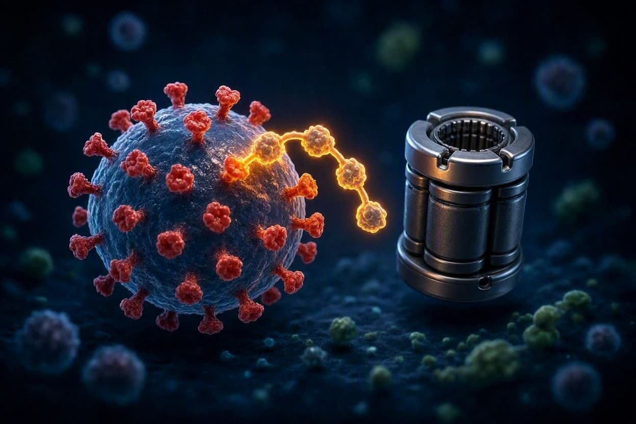
In the silent, microscopic war between pathogens and their hosts, survival often depends not on brute force, but on subtle manipulation. Viruses, lacking the complex machinery of cellular life, must borrow from their hosts to replicate and thrive. Among the most sophisticated tools they exploit is the ubiquitin-proteasome system, a cellular quality-control mechanism that normally tags damaged or unnecessary proteins for destruction. Recent research highlights how certain viruses have evolved to hijack this very system, turning a defensive cleanup crew into an accomplice for immune evasion.
The ubiquitin-proteasome pathway is essential for maintaining cellular health, regulating everything from cell cycle progression to immune responses. By attaching small ubiquitin molecules to specific proteins, cells mark them for degradation by the proteasome, a large protein complex that acts as the cell’s shredder. This process is crucial for presenting viral antigens to the immune system, allowing T-cells to recognize and eliminate infected cells. However, some viruses have developed strategies to interfere with this tagging process, effectively hiding from the immune surveillance that relies on it.
Scientists have identified specific viral proteins that mimic or interact with host enzymes involved in ubiquitination. These viral factors can either prevent the tagging of viral proteins, allowing them to accumulate and replicate, or they can accelerate the degradation of host immune signaling molecules. For instance, some viruses target key components of the interferon signaling pathway, ensuring that the alarm bells of the immune system are silenced before they can ring out across the cellular neighborhood.
This molecular mimicry is a testament to the evolutionary pressure exerted by the host immune system. Over millennia, viruses that could subtly manipulate the ubiquitin-proteasome system gained a significant survival advantage. The result is a delicate balance where the virus must disrupt just enough of the host’s regulatory network to evade detection without causing immediate cell death, which would halt its own replication cycle. It is a dance of precision, where too much disruption leads to host collapse, and too little leads to viral clearance.
Understanding these mechanisms has profound implications for antiviral therapy. Traditional approaches often target viral replication enzymes, but resistance can develop quickly. Targeting the host-virus interface, particularly the interactions within the ubiquitin-proteasome system, offers a new avenue for treatment. By developing small molecules that block viral hijacking of this pathway, researchers hope to restore the cell’s natural ability to detect and destroy the invader.
Recent studies have focused on identifying specific E3 ubiquitin ligases that are co-opted by viruses. These enzymes are responsible for selecting which proteins get tagged with ubiquitin. By inhibiting the interaction between viral proteins and these host ligases, it may be possible to prevent the degradation of critical immune sensors. This approach requires a deep understanding of the structural biology involved, as the interactions are often transient and highly specific.
The complexity of the ubiquitin-proteasome system means that any therapeutic intervention must be carefully calibrated to avoid disrupting normal cellular functions. Since this pathway regulates many essential processes, broad inhibition could lead to severe side effects. Therefore, the goal is to design drugs that are highly selective, targeting only the viral-mediated disruptions while leaving the host’s natural regulatory mechanisms intact.
As we deepen our understanding of these molecular interactions, we move closer to therapies that work with the body’s natural defenses rather than against them. The viral hijacking of the ubiquitin-proteasome system is a reminder of the intricate connections within biological systems, where even the mechanisms of destruction can be repurposed for survival.
AI Image Disclaimer: The visual representations included here are AI-generated illustrations intended to conceptualize molecular interactions and are not actual scientific micrographs.
Sources: Nature Reviews Microbiology Cell Host & Microbe Science Daily
Published by Banx Network. This article is part of the Banx decentralized media programme, powered by the BXE token on the XRP Ledger.