In forests, fields, and city gardens, ants move with a purpose that is easy to overlook. Their paths weave through soil and stone, beneath leaves and along the quiet seams of the ground. Each step is small, almost weightless, yet together they form one of the most enduring presences in the natural world.
To watch an ant is to glimpse a fragment of a much larger story—one that stretches across continents and millions of years. Scientists estimate that thousands of ant species inhabit the Earth, each with its own subtle variations of shape, size, and behavior. Yet for researchers trying to understand this diversity, even the smallest detail can matter.
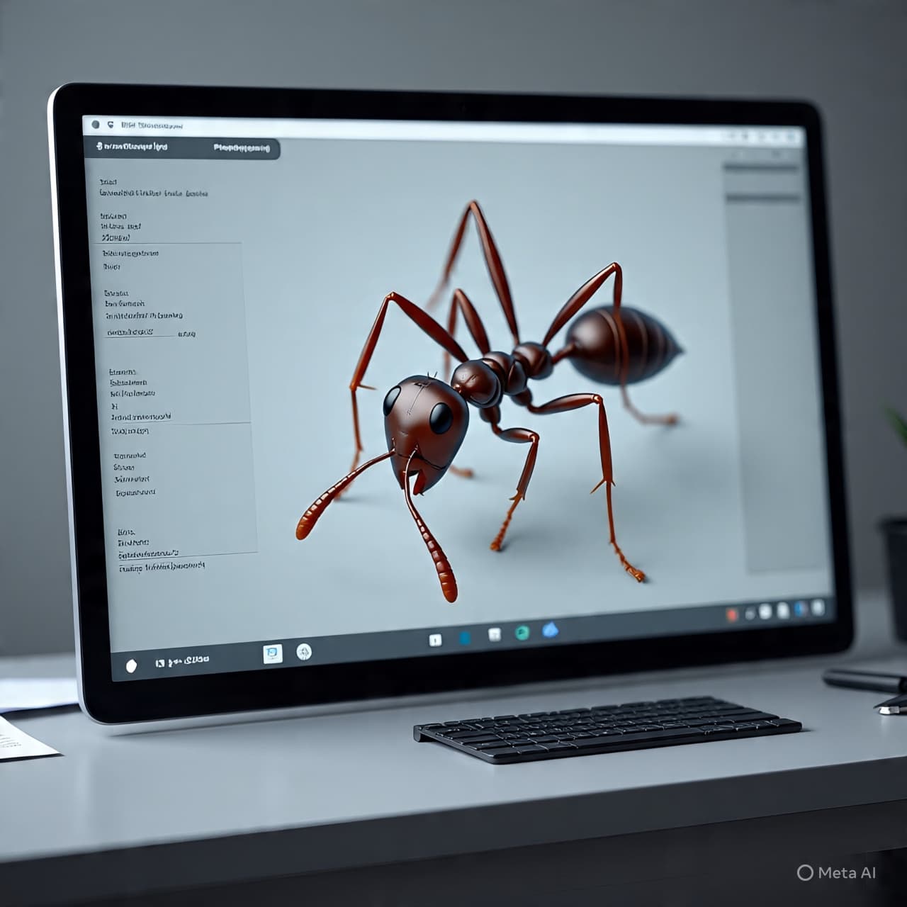
Recently, a group of scientists turned to a new method of observation: rapid three-dimensional scanning. With specialized imaging systems capable of capturing extremely fine structural detail, they began creating digital models of ants preserved in research collections.
The result is the beginning of a digital library containing high-resolution 3D models of more than 800 ant species. Each specimen is scanned from multiple angles, allowing computers to reconstruct a precise representation of the insect’s body—its antennae, joints, mandibles, and delicate segments—down to microscopic features.
For taxonomists, these details are essential. The classification of ants often depends on minute differences in anatomy: the curvature of a thorax, the proportion of leg segments, the subtle shape of a mandible. Traditionally, studying these features has required access to physical specimens stored in museum collections, sometimes scattered across institutions and continents.
Three-dimensional imaging offers another possibility. Once scanned, a specimen can be explored digitally, rotated on screen, enlarged many times beyond its natural size, and examined by researchers anywhere in the world.
The scanning process itself unfolds quickly. Using high-speed imaging systems similar to micro-computed tomography, scientists capture thousands of images in rapid succession as the specimen rotates within the instrument. These images are then assembled into a volumetric model, preserving the insect’s structure with remarkable precision.
Such models do more than replicate appearance. They allow scientists to measure structures directly, compare species in new ways, and build datasets that can be analyzed computationally. Over time, these digital archives may help reveal patterns of evolution, adaptation, and geographic distribution across the enormous diversity of ant life.
There is also a quieter dimension to the effort. Natural history collections hold millions of specimens gathered over centuries of exploration. Many are fragile, and some represent species found only rarely in the wild. Digitizing them creates a form of preservation—an additional layer of record that remains accessible even as the physical specimens age.
The ants themselves remain unchanged, their bodies resting in museum drawers or research cabinets. Yet through scanning and digital reconstruction, they begin to move again—this time across computer screens and databases, accessible to scientists, students, and curious observers across the world.
The study reports that researchers used rapid 3D imaging techniques to digitize more than 800 ant species, creating a detailed virtual library intended to support taxonomy, evolutionary research, and global access to biological collections.
AI Image Disclaimer
Illustrations were created using AI tools and are not real photographs.
Source Check
Credible coverage and/or primary reporting exist from: Nature Communications ScienceDaily Phys.org Smithsonian Magazine The Guardian
Published by Banx Network. This article is part of the Banx decentralized media programme, powered by the BXE token on the XRP Ledger.