
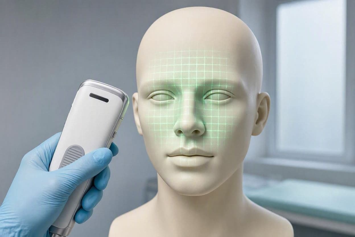
The human face is a landscape of identity, expressing emotion, heritage, and health. In medicine, capturing the precise geometry of this landscape is crucial for diagnosis, treatment planning, and reconstructive surgery. Traditionally, this has required bulky, expensive equipment confined to specialized clinics. Now, a new handheld device using structured light technology offers a portable alternative, bringing high-precision 3D imaging to the point of care. This advancement invites reflection on how technology can democratize access to detailed medical data, making healthcare more flexible and patient-centered.
Structured light works by projecting a pattern of light onto the face and analyzing how it deforms over the contours of the skin. This method allows for rapid, non-invasive capture of three-dimensional data. The clinical validation of a handheld device means that it has been tested against gold-standard systems to ensure its accuracy and reliability. For clinicians, this validation provides the confidence needed to integrate the tool into routine practice.
The portability of the device is a significant advantage. It can be used in bedside settings, outpatient clinics, or even remote locations where large scanners are unavailable. This flexibility is particularly valuable for patients who have difficulty moving or for pediatric cases where minimizing stress and movement is essential. It transforms the imaging process from a logistical challenge into a simple, quick procedure.
In fields like craniofacial surgery, orthodontics, and dermatology, precise measurements are vital. Tracking changes in facial structure over time helps monitor disease progression or treatment outcomes. The handheld device allows for frequent, consistent scans, creating a detailed longitudinal record. This data supports more informed decision-making and personalized care plans.
The validation study likely involved comparing the device’s measurements with those from established stationary scanners. Results showing high correlation would confirm that the handheld unit does not compromise quality for convenience. Such evidence is critical for regulatory approval and adoption by healthcare providers who rely on precision for surgical planning.
Beyond clinical utility, the technology enhances patient comfort. Traditional scanning can be intimidating, requiring patients to remain still in a fixed position for extended periods. A handheld device is less intrusive, allowing for a more natural interaction. This human-centric design improves the overall experience, reducing anxiety and increasing cooperation.
The implications for telemedicine are also promising. With accurate 3D data captured locally, specialists can remotely assess patients, providing expert opinions without the need for travel. This capability expands access to specialized care, particularly for underserved communities. It bridges the gap between local providers and global expertise.
As the technology matures, integration with artificial intelligence could further enhance its value. Algorithms might automatically detect anomalies or suggest treatment options based on the 3D models. This synergy between hardware and software promises a future where diagnostic tools are not just measuring devices but intelligent assistants.
The clinical validation of a handheld structured light device for 3D facial morphometrics marks a step forward in portable medical imaging. By combining accuracy with convenience, the technology supports better patient care in various clinical settings, enhancing both diagnostic precision and comfort.
AI Image Disclaimer: The images associated with this article are AI-generated visualizations intended to symbolize medical imaging and facial analysis, using respectful and abstract imagery.
Sources: Journal of Medical Imaging Health Technology Assessment Reports Clinical Dermatology Journals
Published by Banx Network. This article is part of the Banx decentralized media programme, powered by the BXE token on the XRP Ledger.