
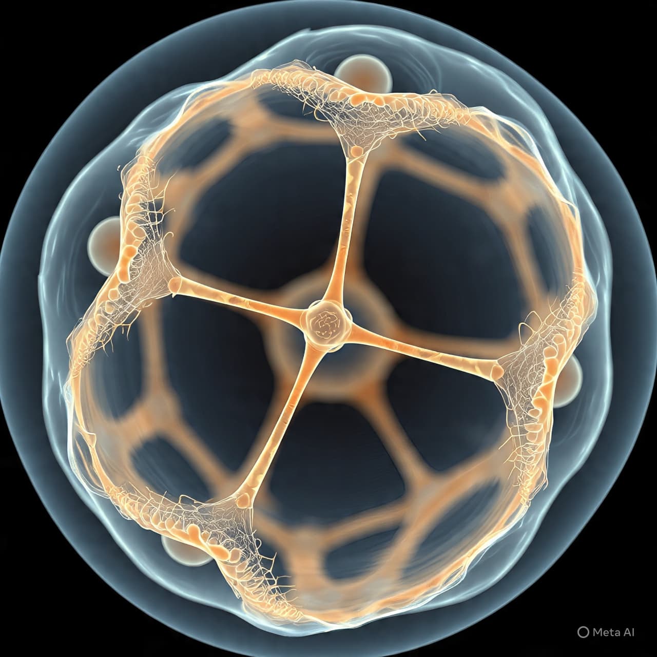
Imagine, for a moment, the intricate artistry of a spider weaving its web. Each strand, though delicate, is woven with purpose, working together to create a structure that holds the potential for something much greater. This act of weaving, though seemingly spontaneous, is governed by an unseen choreography — a dance between elements that form a greater whole. In the microscopic world, cells do something very similar when it comes to the creation of an egg. Like an artist assembling a masterpiece, they use a dynamic scaffold of proteins, namely actin and microtubules, to create the complex structure that will eventually give rise to life. This process, though unseen by the human eye, is a profound example of the elegance and precision with which nature operates. But how exactly do cells orchestrate this delicate construction? The answer lies in the coordinated efforts of two fundamental components: actin filaments and microtubules.
The process of egg formation, or oogenesis, is a marvel of cellular engineering. It begins with the fundamental building blocks of the cell: actin filaments and microtubules. These two components, though distinct in their structure and function, work in tandem to build the architecture of the egg cell, each playing a vital role in ensuring the egg’s proper formation.
Actin filaments are long, thin protein fibers that provide structural support to the cell. Think of them as the scaffolding of a building — without them, the cell would lose its shape and its ability to perform the complex tasks required during egg development. Actin filaments are dynamic, constantly being assembled and disassembled in a delicate balance. This fluidity allows the cell to adjust its shape and structure as needed, guiding the egg through various stages of its formation.
Microtubules, on the other hand, are slightly more rigid structures, resembling the beams of a building’s frame. These tubes, made from tubulin proteins, are responsible for transporting key components within the cell. They form an internal highway system, ensuring that critical molecules, including proteins and organelles, are delivered to the right locations during egg development. They also play a crucial role in the cell’s division, a process essential for egg maturation.
In oogenesis, actin and microtubules work in harmony to establish the egg’s correct form. During the early stages of egg development, actin filaments guide the organization of the cell’s components, ensuring that the egg is the proper size and shape. As development continues, microtubules come into play, arranging and sorting the contents within the egg. The two components work together, ensuring that each molecule and structure is placed in precisely the right location.
The synergy between actin and microtubules is not limited to the physical structure of the egg. These elements also influence how the egg responds to external signals, such as hormonal cues, that regulate its development. The proteins that make up these filaments can interact with one another, coordinating responses to these signals. This cellular choreography ensures that the egg’s development is tightly regulated and occurs at the right pace, with everything in place when it’s time for fertilization.
The process of egg formation is a brilliant display of cellular coordination. Through the combined efforts of actin and microtubules, the cell creates the complex scaffold needed for egg development — a scaffold that will ultimately support the creation of new life. This intricate dance of molecular components is a testament to the elegance of nature’s design, where even the smallest structures have profound roles in the grander narrative of life. As we continue to study the inner workings of cells, we gain deeper insight into the mechanisms that make life possible. And in this delicate balance, we find a story of cooperation and purpose that echoes through the very fabric of existence.
AI Image Disclaimer: "Images in this article are AI-generated illustrations, meant for concept only." "Visuals are created with AI tools and are not real photographs." "Illustrations were produced with AI and serve as conceptual depictions." "Graphics are AI-generated and intended for representation, not reality." Sources: Nature Science Daily Cell The New York Times National Institutes of Health (NIH)
Published by Banx Network. This article is part of the Banx decentralized media programme, powered by the BXE token on the XRP Ledger.