
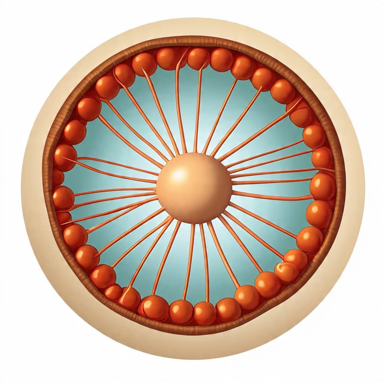
In the quiet interior of a cell, construction happens without noise. There are no cranes or blueprints, no foremen calling out instructions. And yet, again and again, an egg cell—one of the largest and most consequential cells in the body—takes shape with astonishing precision. Its form is not accidental. It is built.
For decades, biologists described the egg as a vessel: a container for genetic material and nutrients, waiting passively for fertilization. But recent research has shifted that image. The egg is not merely filled; it is assembled. Its architecture emerges from a dynamic collaboration between two internal structures—actin filaments and microtubules—that together form a living scaffold, shaping the cell from within.
Actin is often associated with motion. In muscles, it contracts; in migrating cells, it pushes membranes forward. Microtubules, by contrast, are rigid and directional, acting as highways for cargo and organizers of internal space. In egg cells, these systems do not operate in isolation. They interlock, respond to one another, and divide labor in ways that give the egg its size, polarity, and internal order.
As an egg grows, actin filaments create a flexible mesh beneath the cell membrane. This network helps the cell expand without tearing, distributing tension evenly as volume increases. At the same time, microtubules extend inward like spokes, guiding the transport of proteins, RNA, and organelles to precise locations. The egg’s interior becomes zoned: some regions rich in nutrients, others primed for early development.
What makes this process remarkable is its coordination. Actin can influence where microtubules form, while microtubules can reshape actin networks in return. Signals pass back and forth, ensuring that growth remains symmetrical—or deliberately asymmetrical, depending on the species. In many animals, this asymmetry determines the future body plan of the embryo, long before fertilization occurs.
This internal scaffolding is not static. It rearranges constantly as the egg matures. Portions stiffen, others relax. Structures appear, dissolve, and reappear elsewhere. The egg is less like a finished building than a construction site that never quite closes, adjusting itself in anticipation of what comes next.
Understanding this choreography matters beyond basic biology. Errors in cytoskeletal coordination can lead to failed egg development or infertility. Similar actin–microtubule interactions also shape neurons, immune cells, and cancer cells, making the egg a powerful model for how large, complex cells organize themselves.
Seen this way, the egg is not just the beginning of life but a demonstration of cellular engineering at its most elegant. Without eyes or intent, the cell measures space, balances forces, and builds a structure capable of becoming something vastly more complex. Before life divides and differentiates, it first learns how to hold itself together.
AI Image Disclaimer
Illustrations were created using AI tools and serve as conceptual representations.
Sources
Nature Science Cell Proceedings of the National Academy of Sciences Developmental Cell
Published by Banx Network. This article is part of the Banx decentralized media programme, powered by the BXE token on the XRP Ledger.